For hundreds of years, the human cadaver has been a critical tool for medical teaching, but it’s been problematic for reasons as diverse as cost, transport, storage, spiritual beliefs or just general queasiness.
Monash University in Australia might finally have the answer to a majority of these obstacles: The first commercially available kit of realistic, full-color body parts produced by a 3D printer.
A paper from Monash University titled “The Production of Anatomical Teaching Resources Using Three-Dimensional Printing Technology” lists several advantages of using 3D printed cadavers, including “accuracy, ease of reproduction, cost-effectiveness and the avoidance of health and safety issues associated with wet fixed cadaver specimens or plastinated specimens.”
Looking inside the body
Specimens are printed by Monash using 3D Systems ColorJet Printing (CJP) technology. The ProJet series of color printers are easy to use. Most importantly, they produce models in the exact colors that Monash needs for realistic 3D printed body parts.
“The full color is essential to reproducing a combination of realistic color fidelity and ‘coding’—vessels in red or blue, nerves in yellow, for example—that is valuable in teaching,” says Paul McMenamin, director of the Centre for Human Anatomy Education (CHAE) at Monash University.
McMenamin believes his team’s simple and cost-effective anatomical kit could dramatically improve knowledge for medical students and practicing doctors. It could even contribute to better surgical outcomes for patients.
“For centuries cadavers bequested to medical schools have been used to teach students about human anatomy, a practice that continues today,” says McMenamin. “However, many medical schools report either a shortage of cadavers or find their handling and
storage too expensive as a result of strict regulations governing where cadavers can be dissected.
“We believe our kit will revolutionize learning for medical students by enabling them to look inside the body and see the muscles, tendons, ligaments and blood vessels. At the moment it can be incredibly hard for students to understand the three-dimensional form of human anatomy, and we believe this kit will make a
huge difference.”
Realizing an ‘ah ha’ moment
Cadavers printed in 3D might seem like a logical progression for the medical community, but it took technological progress in 3D printing to make it happen. The 3D Systems machines used by Monash University deliver the ability to print full-color models at relatively high speeds at a cost that provides a marked improvement over plastic models or plastination of human remains.
“I was looking for a way to produce more anatomy prosections and maybe plastinate them, but realized it would take decades and more than a half-million dollars to set up a plastination lab,” says McMenamin. “Each specimen would have to be dissected and prepared and then I would have one of that specimen.
“So we thought ‘why don’t we scan them (CT or laser), make color STL or VRML files, and print them so we can make lots of copies’. Seems obvious now, but it was sort of an ‘ah ha’ moment.”
Thanks to the 3D Systems printers, Monash University can produce parts that range from a full body to head and neck, upper limb, pelvis and lower limb, and thoracic and abdominal regions. A deal with German anatomical model makers Erler-Zimmer makes the cadavers available for purchase online, with delivery within weeks at a fraction of the cost of an embalmed or plastinated body.
The Monash series also includes anatomically correct models that would be impossible to visualize in an embalmed body – such as 3D prints of the vasculature of the brain with fine veins and arteries embedded within the skull.
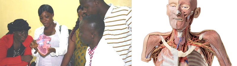
Making a difference in Liberia
A recent project showed just how much of a difference a 3D printed cadaver can make to a university in need — in this case, the University of Liberia’s Dagliotti Medical School.
Inspired by a speech by Dr. Ian Crozier, a doctor who had contracted Ebola while working in Sierra Leone, McMenamin arranged for a full set of 3D prints and a set of posters of histological (a microscopic anatomy of cells and tissues) images to be sent to the school.
McMenamin also volunteered his time to teach faculty and students how to use the 3D anatomy kit. His accommodations and logistical support in Liberia were provided by ACCEL (Academic Consortium Combating Ebola in Liberia), an effort led by the University of Massachusetts Medical School and funded by Paul G. Allen’s #TackleEbola initiative.
In exchange for his donations and teaching, McMenamin has the satisfaction of helping a desperately poor and understaffed medical school provide better anatomical teaching for a new generation of Liberian doctors.
“Helping the medical school in Liberia with the support of my CHAE team and Monash University has been the best thing I have done for my fellow human beings,” says McMenamin. “The students there were just so grateful for any help that was provided. It was very humbling.”
McMenamin is likely to have more achievements in the near future about which to be humble: Using the latest 3D printing technologies from 3D Systems, his team is working on interactive, dissectible 3D anatomical reproductions that could be used to help train future surgeons.
Thanks to McMenamin and 3D printing, the cadaver, in all its full-scale and full-color glory, is gaining a new lease on life in medical universities around the world.
http://www.3dsystems.com/sites/www.3dsystems.com/files/cs_3ds_monash_cadaver_030916.pdf

